Introduction
Bronchiectasis is a medical term composed of two Greek words: “bronkos” meaning “breathing tube” and “ektasis” meaning “stretching” or “extension”.
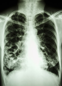
This image of bronchiectasis (thanks to www.nhlbi.nih.gov for this image) shows that there is usually an accumulation of several bronchiectasis. These dilatations of abnormal bronchial tubes are due to a loss of the muscular and elastic layers of the chronically infected bronchial tubes within one of the lung segments.
Chronic bronchitis in longterm smokers often leads to such destruction and multiple bronchiectasis. However, bronchiectasis can also be found in the upper lobes of the lungs, which is typical for tuberculosis. Another disease that is associated with bronchiectasis is cystic fibrosis (a congenital lung disorder with a lack of surfactant production), where bacterial infections are common.
Viral illnesses such as measles, pertussis and influenza in unimmunized people can cause bronchiectasis following one of these viral diseases.
Signs and symptoms
Chronic cough, fever, weight loss and the chronic production of secretions that look like pus are all associated with bronchiectasis. This pussy mucous comes from the dilated diseased bronchial tubes, which are infected with problem bacteria like Pseudomonas aeruginosa, Staphylococcus aureus and Haemophilus influenzae, all of which are very difficult to treat successfully with antibiotics. Bronchiectasis often is associated with a chronic lack of oxygen in the system and this leads to clubbing of the fingers (thanks to www.indianpediatrics.net for this image). Coughing up of blood (=hemoptysis) is also commonly found.
Diagnostic tests
Chest X-rays show characteristic signs such as the “toothpaste lines” and “tram tracks” or “tram lines”, which when present are characteristic of bronchiectasis. However, the details are much more visible (in almost all bronchiectasis cases) with the a lung CT scan, particularly the high resolution CT scan (=HRCT). In some cases the respirologist may decide to do a bronchoscopy, just to rule out other pulmonary diseases such as lung cancer, congenital abnormalities, tuberculosis or to obtain a good sample of pus right from where the source is inside the bronchial tube affected by a bronchiectasis. The physician likely will order several samples of sputum and send this for laboratory analysis with culture and sensitivity testing to a battery of antibiotics. This way the physician can be guided by the results of the best fit of antibiotic activity against a particular bacterial strain isolated.
Treatment
Treatment in the earlier cases of bronchiectasis is directed against the chronic infection in an attempt to prevent further deterioration of the condition and to prevent pneumonia. Broad spectrum antibiotica are used such as amoxicillin, tetracycline, or trimethoprim-sulfamethoxazole, if a mixed bacterial flora is shown to be present on cultures from the coughed up secretions (=sputum cultures). However, often there are problem bacteria present with flare-ups such as Pseudomonas aeruginosa and special antibiotics such as ticarcillin, gentamycin, tobramycin, ceftazidime, and ciprofloxacin have to be used for a period of time. If there is an underlying chronic bronchitis problem that has caused the bronchiectasis many physicians use one of the broad spectrum antibiotics to keep the infective process under control (prophylactic antibiotic), but will do sputum cultures with flare-ups from time to time and switch the antibiotics to one, which will be effective against the particular bacterium found at that time. Other physicians do not want to use prophylactic antibiotics for fear of creating “superbugs” that can be resistant against most or all known antibiotics (Ref. 4, page 1331). Along with the antibiotic therapy chest physiotherapy is given and can be learnt by family members and be administered at home. Clapping of the chest wall, postural drainage and deep breathing exercises are all part of this regime. By draining the secretions from the lungs bacteria have less fluid on which to grow.
Surgery
If the bronchiectasis condition is confined to one or two lung segments, surgical removal of this segment can be considered. The mortality in most centers in the US is now 1% to 2% in the hands of a chest surgeon. Considering how sick these patients are, this is considered low. However, surgery can only be contemplated, if the bronchiectasis condition is confined to one area of a lung. In cases where there is an acute coughing up of blood (called “hemoptysis”), the chest surgeon may have to remove a particularly bad area of the lung where a bronchial artery has been eroded from a pus pocket of a bronchiectasis.
Prevention
The key as in many medical illnesses is prevention of bronchiectasis. To avoid cigarette smoking will prevent chronic bronchitis, one of the major causes of this disease. Immunization against measles, pertussis and influenza prevents major damage to the bronchial tubes. Any bacterial infection of the airways must be treated aggressively with antibiotics in order to prevent the erosion of the muscular and elastic layers of the bronchial tube, which is at the core of the development of bronchiectasis.
References:
1. Noble: Textbook of Primary Care Medicine, 3rd ed., Copyright © 2001 Mosby, Inc.
2. National Asthma Education and Prevention Program. Expert Panel Report II. National Heart, Lung and Blood Institute, 1997.
3. Rakel: Conn’s Current Therapy 2002, 54th ed., Copyright © 2002 W. B. Saunders Company
4. Murray & Nadel: Textbook of Respiratory Medicine, 3rd ed., Copyright © 2000 W. B. Saunders Company
5. Behrman: Nelson Textbook of Pediatrics, 16th ed., Copyright © 2000 W. B. Saunders Company
6. Merck Manual: Bronchiectasis (thanks to http://www.merckmanuals.com for this link).
7. Goldman: Cecil Textbook of Medicine, 21st ed., Copyright © 2000 W. B. Saunders Company
8. Ferri: Ferri’s Clinical Advisor: Instant Diagnosis and Treatment, 2004 ed., Copyright © 2004 Mosby, Inc.
9. Rakel: Conn’s Current Therapy 2004, 56th ed., Copyright © 2004 Elsevier


