
Introduction
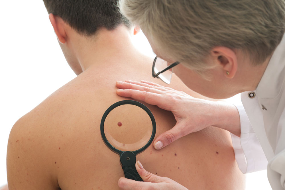
In general, one of the important tasks of a physician and dermatologist is to diagnose moles and skin cancer.
Indeed, a lot of time is spent at continuing education conferences to train the eye of physicians by showing hundreds of slides of different presentations of benign skin lesions and cancerous skin lesions. To repeat, it is important to distinguish the good (benign skin lesion) from the ugly (malignant).
Certainly, I can only give a two dimensional overview of a clinical problem, which is three dimensional. In detail, there is a fourth dimension by way of the fact that with development over time the good skin tumor does not change, but the ugly tumor (melanoma and skin cancer) takes off and metastasizes. For this reason, if you see a skin lesion on your body that resembles in the slightest a cancerous tumor (see picture link below), see your physician right away.
Provided that your physician cannot identify what lesion it is, you should be referred to a skin specialist (=dermatologist), because that is what this professional specializes in. On the contrary, too many people are still ignorant about the early cancerous skin lesions on their skin and too many people still die needlessly of them.
Moles and skin cancer
Signs and symptoms
Below are links to sites that show pictures of skin moles. Depending on what type of skin mole there is (flesh color, brown, red, black) and from what tissue component of the skin it originates from, there are a great number of different skin lesions, which can be divided into benign and malignant.
Common skin moles and skin cancer (a small selection)
_________________________________________________________________
Benign skin lesions
They are well rounded, usually soft, contain one normal cell type; there is no or very slow local growth; never produces metastases
- junction nevus : initially flat, later slightly raised brown or black benign skin lesion
- compound nevus : flat periphery, elevated center in mostly pigmented lesion; resembles melanoma, sometimes only biopsy can decide
- dermal nevus : dome shaped pigmented cell nevus; with a lot of vessels might resemble basal cell carcinoma
- seborrheic keratosis: a warty dermal nevus type, can look deep black, but is harmless, can be treated by liquid nitrogen; delineate from melanoma!
- hemangiomas : often raised, red; laser therapy often useful
- actinic keratosis : a pre-malignant lesion in sun exposed areas; if not removed, turns into basal cell carcinoma (a form of skin cancer)
Malignant skin lesions
They are irregularly shaped, hard, consist of one malignant cell clone that multiplies rapidly after an initial slower growth period; cell clusters invade distant body parts (called “metastases”)
- melanoma : usually deeply pigmented (brown, black), but can also be flesh colored; once it invades more than 6 mm into the skin, it forms metastases rapidly leading to death
- skin cancer : basal cell cancer (thanks to www.cbc.ca for image) and squamous cell cancer (thanks to commons.wikimedia.org for this image) are the commonest, but there are 29 different types
_________________________________________________________________
Treatment
Surely, this is a very important topic, but one where you need the help of a physician and possibly a specialist like a dermatologist. Specifically, I have described skin biopsies under both melanoma and skin cancer. One elegant method is Mohs’ surgery, which can get results of almost 100% cure rates in an early stage of skin cancer.
Decisions have to be made who needs liquid nitrogen, who needs surgical excision for diagnostic and possibly curative purposes and who needs the more involved Mohs’ surgery. Truly, your family doctor will coach you through this complex field of medicine. Sometimes a dermatologist has to help as well.
More info on skin cancer and moles:
American Academy of Family Physicians about early melanoma detection (thanks to www.aafp.org for this link).
References
1. Habif: Clinical Dermatology, 3rd ed.,1996, Mosby-Year Book, Inc.
2. The Merck Manual, 7th edition, by M. H. Beers et al., Whitehouse Station, N.J., 1999. Chapter 117.
3. Cotran: Robbins Pathologic Basis of Disease, 6th ed.,1999, W. B. Saunders Company
4. Noble: Textbook of Primary Care Medicine, 3rd ed., 2001, Mosby, Inc.
5. Rakel: Conn’s Current Therapy 2001, 53rd ed., 2001, W. B. Saunders Company
6. Goroll: Primary Care Medicine, 4th ed., 2000, Lippincott Williams & Wilkins
7. Richard J. Lewis, M.D. at the 42nd Annual St. Paul’s Hosp. CME Conf., Nov.1996, Vancouver/BC
8. Jerry Shapiro, Prof. Dermatol., UBC, at 45th Annual St. Paul’s Hosp. CME Conf., Nov.1999, Vancouver/BC
9. D Seager Int J Cosmet Surg Vol 6, No. 1, 1998: 27-31.
10. Townsend: Sabiston Textbook of Surgery, 16th ed.,2000, W. B. Saunders Company
11. Ferri: Ferri’s Clinical Advisor: Instant Diagnosis and Treatment, 2004 ed., Copyright © 2004 Mosby, Inc.
12. Rakel: Conn’s Current Therapy 2004, 56th ed., Copyright © 2004 Elsevier


