Hemolytic anemias from changes in the RBC’s can occur when the red blood cell membrane differs the regular red blood cells built. This is what is affected in the hereditary conditions of hereditary spherocytosis or hereditary elliptocytosis. A page regarding “hereditary elliptocytosis and spherocytosis” is linked from “Related Topics” below.
Another hereditary enzyme defect can lead to hemolytic anemias from changes in the RBC’s. This time the genetic defect is in the heme synthesis of the red blood cell; this can present in infancy when even after a short exposure to sunlight there is a burning pain in the skin. This form of anemia is called erythropoietic protoporphyria and this can be accessed through “Related Topics” below, where this condition is dealt with in more detail.
Here is a brief review how hemolytic anemias from changes in the RBC’s develop due to genetic changes in the red cell membrane.
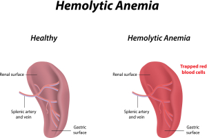
In spherocytosis the surface of the red cell membrane is diminished compared to normal red cells and this results in less flexibility. When the spherocytes (abnormally round red cells) are filtered though the capillaries of the spleen, they get destroyed and hemolysis occurs. Anemia ensues.
With elliptocytosis the cytoskeleton of the red blood cell is weakened resulting in elliptical deformities, called elliptocytes. In the spleen these cells are removed and destroyed, often leading to an enlarged spleen.
With erythropoietic protoporphyria it is the heme metabolism, which leads to hemoglobin synthesis that is interrupted by the congenital disorder. The majority of cases are due to an autosomal recessive enzyme deficiency for the synthesis of the heme portion of hemoglobin; these cases have minor photosensitivity. However, the other, smaller subgroup have another defect in the heme synthesis, which is X-linked and they have more severe photosensitivity as well as liver damage.
These are examples where relatively minor defects in the metabolism of one cell group (the red blood cell formation in the bone marrow)has a far-reaching impact on these patients. It can lead to enlarged spleen first, in more severe cases also an enlarged liver. It can lead to pigmented gallstones in the X-linked erythropoietic protoporphyria as well as chronic liver disease and liver cirrhosis.
References:
1. The Merck Manual: Hemolytic anemia
2. Noble: Textbook of Primary Care Medicine, 3rd ed., Mosby Inc. 2001
3. Goldman: Cecil Medicine, 23rd ed., Saunders 2007: Chapter 162 – APPROACH TO THE ANEMIAS


