Introduction
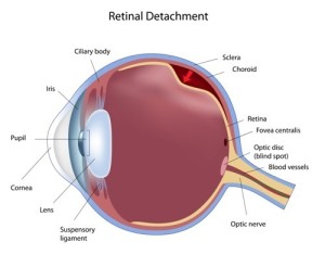
Notably, retinal detachment means that the retina, which is like a wall paper lining in the back of the eye, gets detached. It is important to realize that this is a medical emergency as an eye specialist has to repair this condition as soon as possible to prevent permanent blindness. Certainly, retinal detachment can occur as a result of a tear or as a result of traction from scarring or proliferative retinopathy. It is important to realize that an underlying tear could be from myopia, could be following trauma to the eye or after cataract surgery. Truly, retinal detachment from traction commonly occurs with proliferative diabetic retinopathy. However, retinal detachment can develop also following severe cases of uveitis, or tumors in the choroidal layer behind the retina. It is here that the blood vessels are located.
Signs and Symptoms
There is no pain with this condition. Initially there might be dark floaters or flashes of light. Suddenly there might be a lack of vision following an experience that a “curtain dropped down”.
Diagnostic Tests
The eye specialist uses direct and indirect ophthalmoscopy to evaluate this complex three-dimensional occurrence. B-scan ultrasonography helps the eye specialist to locate the detachment and to delineate the size of it.
Treatment
Depending on what the eye surgeon finds, there are a number of procedures available for the repair of a retinal detachment.
Pneumatic retinopexy is a method where the eye specialist introduces a gas bubble into the vitreous body. This pushes the detached retina up against the wall of the eyeball and subsequently the the specialist uses laser surgery of the retina to “spot-weld” it to reduce the probability of a reoccurrence of the detachment.
Vitrectomy is a surgical method that removes the vitreous body.
Ultimately the eye specialist knows best what you need in a specific case.
References
1. The Merck Manual: Detachment of the Retina
2. Ferri: Ferri’s Clinical Advisor: Instant Diagnosis and Treatment, 2004 ed., Copyright © 2004 Mosby, Inc.
3. Rakel: Conn’s Current Therapy 2004, 56th ed., Copyright © 2004 Elsevier


