Introduction
Hypertensive retinopathy develops in patients who had high blood pressure for a long time. Chronic spasm of the smaller arteries, called arterioles cause this condition.
This leads to accelerated hardening of the arteries (arteriosclerosis). This in turn leads to leakage of some atheromatous arterial vessels. On fundoscopy the physician sees them as hemorrhages that look like flames. In addition, there is a lack of blood supply in other areas of the retina (ischemic changes and infarcts).
With leakage of exudate that contain blood proteins and lipids swelling and scarring occurs in the area of sharpest vision. This has the name fovea or macula and any scarring there results in loss of vision. 4 categories of vision loss exist. In the past this was significant as without treatment there would be an enormous difference in survival.
Signs, Symptoms and Diagnostic Tests
Painless and gradual loss of vision in both eyes occurs when physicians neglect to measure blood pressure. The eye specialist looks through an ophthalmoscope or a slit lamp to see details in the back of the eye. In a patient with high blood pressure he sees various degrees of retinal changes. This in turn depends on the grade of severity of hypertensive retinopathy.
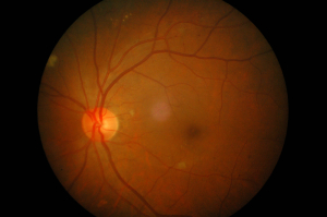
The visual appearance of changes associated with hypertensive retinopathy over the years have received strangely sounding names. There are “cotton wool spots” (mini-infarcts of the retina) and “macular star”. The latter is a ring of exudate from the optic disc to the fovea or macula. This is the area, which normally has the sharpest vision. Such changes severely affect the vision of the person who has this finding. The most awful of all hypertensive retinopathy cases is grade IV retinopathy. This condition involves the optic disc in the edematous process leading to “papilledema”. The reason this is so dangerous is that this condition compresses the central artery and vein simultaneously. This effectively shuts down the circulation to the retina. The finding of papilledema is a medical emergency that requires treatment in an Intensive Care Unit setting.

Hypertensive Retinopathy (60 year old man with diabetes and high blood pressure); click on image to enlarge
Treatment
The treatment is directed at controlling the blood pressure. In the case of “malignant hypertension”, which corresponds with papilledema and grade IV retinopathy, intravenous titration of the blood pressure in an Intensive Care Unit setting might have to be done in an attempt to rescue the patient’s vision. Otherwise the patient will be permanently blind. All other forms of hypertensive retinopathy are treated in the office setting, but close blood pressure control and self blood pressure monitoring at home are an important part of any therapy. Longterm follow-up eye examinations and longterm blood pressure monitoring are important.
References
1. The Merck Manual: Hypertensive Retinopathy
2. Ferri: Ferri’s Clinical Advisor: Instant Diagnosis and Treatment, 2004 ed., Copyright © 2004 Mosby, Inc.
3. Rakel: Conn’s Current Therapy 2004, 56th ed., Copyright © 2004 Elsevier


