
In order to understand the various eye conditions, it is important to develop a general understanding of the “nuts and bolts” of the eye and the adjacent structures. Physicians call this the “anatomy”. I will use various sites that are freely available to you on the Internet to demonstrate the basic principles. This link of the eye anatomy describes how the rays from an image enter the eye and form an image on the back of the eye, called retina. This link of the function of the lens (thanks to www.sapdesignguild.org for this image) describes how through accommodation the rays from an image enter the eye and form an image on the back of the eye, called retina, depending on whether you focus on a near or far target.
As you look at the outer structures of the eye, notice that the eyeball is protected by the eyelids that will close in milliseconds by powerful blinking reflexes when any object threatens to hit the cornea. The outer layer of the eyeball is white (called sclera) and this can be seen around the iris that is colored and is clearly visible through a normal cornea. The opening of the iris is determined by the brightness of the environment (pupil usually wide open in darkness or when in a sympathetic mood, but the pupil is narrow in bright light). The pupil size is also under the influence of drugs where narcotics make the pupil small and tropicamide or phenylephrine makes it wide. Here is another anatomy site that shows how the beam of light gets through the eye lens and the vitreous body to the retina in the back of the eye. It also shows the anterior and posterior eye chambers that are important to know about when it comes to any abnormality of the circulation of the internal eye fluid. When the pressure in this system builds up, glaucoma can develop.
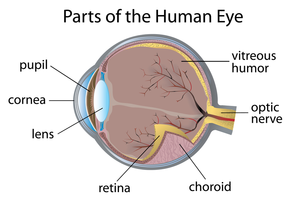
Anatomy Of The Eye (1)
The eye ball is very mobile and this is done through the action of the 6 eye muscles. This link (thanks to webvision.med.utah.edu for this image) tells more about the way how the macula and the rest of the retina are intricately shaped and connected to the optic nerve.
A complex nervous pathway connects the optic nerve impulses to the appropriate part of the brain after the nerve fibers crossed in the optic chiasm. The optic pathways have a very structured way of being processed in the brain as is depicted schematically here (from the Merck Manual) (thanks to www.merckmanuals.com for this link).
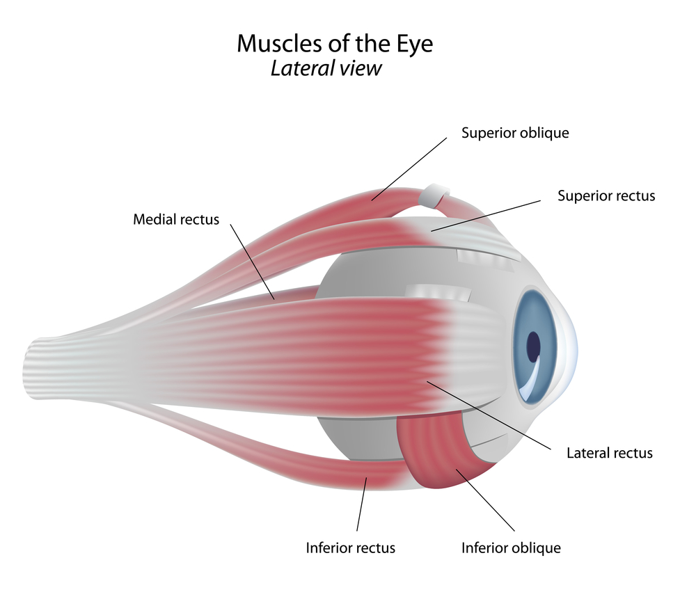
Anatomy Of The Eye (2)
This knowledge is important when it comes to interpreting the location of strokes in certain parts of the brain. A person who develops a stroke in the back part of the brain (occipital area) cannot process the visual images, because the processing unit in the brain is defective. We know from studies with micro-electrodes during brain surgery that the eyes in a case like this are processing images normally (cortical or central blindness).
Other parts of the anatomy of the eye will be further discussed in each of the subchapters.
References:
1. The Merck Manual (thanks to www.merckmanuals.com for this link)
2. Eye conditions: http://www.stlukeseye.com/Conditions/
3. Ferri: Ferri’s Clinical Advisor: Instant Diagnosis and Treatment, 2004 ed., Copyright © 2004 Mosby, Inc.
4. Rakel: Conn’s Current Therapy 2004, 56th ed., Copyright © 2004 Elsevier


