Introduction
The cornea is the front layer of the eyeball as is explained before.
There are several eye diseases and other causes that explain diseases of the cornea. For instance, when an iron piece gets imbedded into the superficial layer of the cornea, which is a common injury in welders, this sets up a traumatic conjunctivitis and the patient will have a lot of pain.
A virus can cause little cornea ulcerations as is the case with herpes zoster, a dangerous corneal condition. Inadequate tear production can also lead to a painful dry eye syndrome (=keratoconjunctivitis sicca). Vitamin A deficiency can lead to a malnutrition state (called “keratomalacia”) of the cornea where it becomes vulnerable to ulcerations with secondary infections. Here is a brief overview of these conditions and their symptoms.
Signs and Symptoms
Various types of corneal problems
| Name (diagnosis): | Symptoms and/or causes: |
| bullous keratopathy | can occur following cataract surgery, related to it is Fuchs’ endothelial dystrophy |
| corneal ulcer | various bacteria and viruses can cause it, photophobia, tearing eyes, pain |
| herpes simplex keratitis | herpes simplex type I can affect the lips and the eyes, see the link for more info |
| herpes zoster ophthalmicus | herpes zoster eye infections can affect various structures of the eye (see links) |
| interstitial keratitis | often associated with uveitis, rare in the US, common in Africa. Syphilis common. |
| keratoconjunctivitis sicca | associated with Sjögren’s syndrome and rheumatoid arthritis. Dry eyes, itchy, not enough tear fluid. |
| keratoconus | inherited condition getting symptomatic at age 10 to 20. Conically shaped cornea. |
| keratomalacia | vitamin A deficiency leads to corneal ulcerations |
| peripheral ulcerative keratitis | often found in rheumatoid arthritis patients. These patients do better when treated with cytotoxic immunosuppression |
| phlyctenular keratoconjunctivitis | occurs in children, seems to be an allergic reaction to an undefined antigen |
| superficial punctated keratitis | a variety of causes can bring on this condition, viral, UV light, contact lenses etc. |
Diagnostic tests
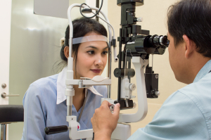
Inspection by the eye specialist and slit lamp examination are the main mode of diagnostic testing. This is helped by bacterial swabs and viral studies, if indicated. Fluorescein staining can help to see superficial defects in the cornea. The Schirmer test shows whether there is enough tear fluid being produced.
Treatment
Treatment is specifically directed against the underlying cause. It is important to stress here that self made home remedies have no place in eye diseases as the patient does not know exactly what is going on. You need a knowledgeable eye specialist who will pinpoint exactly what is going on. Then a treatment plan including regular follow-up visits can be arranged to ensure that the condition gets cured or controlled without permanent loss of vision. The links above contain much information regarding treatment for the various corneal conditions.
References
1. The Merck Manual: Eye diseases
2. Ferri: Ferri’s Clinical Advisor: Instant Diagnosis and Treatment, 2004 ed., Copyright © 2004 Mosby, Inc.
3. Rakel: Conn’s Current Therapy 2004, 56th ed., Copyright © 2004 Elsevier


