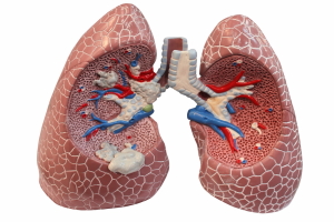
Once a bronchoscopy has confirmed lung cancer, the physician must assess how far the cancer has advanced at the time of diagnosis, before treatment can start. The reason for this are very different treatment protocols that are necessary for the successful treatment of lung cancer.
The lung cancer stage are: stage I, II, IIIA, IIIB and IV. Following the initial X-ray, the physician will then order a CT scan in order to further characterize the lung lesion.
The CT scan can show whether or not the lymph glands in the chest are involved or not. If there are lymph glands in the chest, a thoracic surgeon may be called in to do a mediastinoscopy, where the surgeon can look into the space between the lungs and the rib cage and assess the extent of the metastases in this otherwise difficult to assess space. The oncologist will want to continue to do the staging tests by doing CT scans of the liver, the adrenal glands and the brain to determine whether distant metastases are present. Blood tests and bone scans will rule out bone metastases. Finally, when all this information is gathered, the oncologist can do what is called an ” extent of disease evaluation”. The following would be found for the various stages:
Extent of disease evaluation: Staging of lung cancer
Stage:
I : solitary lung tumor of less than 3 cm (=1 1/4″) in diameter
II : tumor more than 3cm(= 1 1/4″) in diameter, local lymph gland metastases on same side as tumor
IIIA : peripheral lung tumor: invaded chest wall; central lung tumor: invaded distal mediastinal nodes on the same side
IIIB : same as stage IIIA, but more extensive lymph gland invasion involving mediastinal organs and pleural cavity
IV : Any of the above stages, but in addition distal metastases
Why are oncologists “wasting time” to do the staging procedures? Studies over several decades have taught us that treatment of cancer without staging often gives everyone a false sense of security, where they learn later that the real extent of the cancer was much worse than originally thought.
While everyone was thinking no further therapy was necessary, the cancer quietly multiplied and spread until it was too late to do anything about it. With the progress in the treatment of childhood leukemia oncologists learnt that long-term survival and cure rates could show significant improvement with adequate staging in the beginning and by following appropriate treatment protocols. In the last few years this has paid off for lung cancer as well. The following is a survival map for the various stages of lung cancer (modified from Ref. 1 and 2):
Lung cancer (5-year survival rate)
| Stage of lung cancer: | 5-year survival rate: |
| Carcinoma in situ | › 90 % |
| I | 60-80 % |
| II | 25-50 % |
| IIIA | 15-35 % |
| IIIB | › 5 % |
| IV | › 5 % |
Histology of lung cancer
Before starting the treatment the physician will want to know what type of lung cancer he is dealing with. There are two main types, the small cell lung carcinoma (SCLC) and the non-small cell lung carcinoma (NSCLC). About 85% of lung cancers fall into the NSCLC category, 15% belong to the SCLC category. The reason for this distinction is that SCLC cancers respond well to chemotherapy. The bronchogenic carcinoma, which is typically an adenocarcinoma belongs to the NSCLC group. In addition the squamous cell lung carcinoma and the large-cell lung carcinoma also belong into the NSCLC group. Bronchoscopy and histological evaluation of tumor biopsies help to classify the tumor type before a decision is made regarding treatment.
References:
1. Cancer: Principles &Practice of Oncology, 4th edition, volume 1. Edited by V.T. De Vita,Jr.,et. al J.B. LippincottCo.,Philadelphia, 1993.Chapter on lung cancer.
2. Cancer: Principles&Practice of Oncology. 5th edition, volume 1. Edited by Vincent T. DeVita, Jr. et al. Lippincott-Raven Publ., Philadelphia,PA, 1997. Chapter on lung cancer.
3. The Merck Manual, 7th edition, by M. H. Beers et al., Whitehouse Station, N.J., 1999. Chapter 81: Tumors of the lung.
4. GW Krystal et al. Clin Cancer Res 2000 Aug;6(8):3319-3326.
5. BJ Druker et al. N Engl J Med 2001 Apr 5;344(14):1031-1037.
6. MJ Mauro et al. Curr Oncol Rep 2001 May;3(3):223-227.
7. Conn’s Current Therapy 2004, 56th ed., Copyright © 2004 Elsevier
8. Ferri: Ferri’s Clinical Advisor: Instant Diagnosis and Treatment, 2004 ed., Copyright © 2004 Mosby, Inc


