
In the first place the physician must do staging of the cancer of the uterus first before embarking on any cancer treatment. This is like with all the other cancers, where we saw how important it is to complete all the above mentioned tests first prior to treatment. When these tests are complete, the physician can individualize treatment according to the stage the cancer is in for the best long-term results.
Sometimes a woman may be irate with the specialist as the diagnosis may already be known from the histology of the dilatation and curettage (uterine cancer), but the cancer stage has yet to be determined. The specialist needs to explain to the woman that depending on what stage is found the treatment can be radically different and this is the reason why there is no room for short-cuts.
Staging in the past
In the past, particularly with uterine cancer the work-up was not that thorough often neglecting to look for possible pelvic metastases. As a result a simple hysterectomy was done leaving pelvic or paraaortic lymph glands untreated. Of course, this led to very disappointing survival data. With proper investigation this would never have happened. Cancer is very unforgiving and there is no room for errors.
The revised FIGO staging (1988, slightly modified in 2000) is what is used in the major Cancer Clinics throughout the U.S.:
Staging of uterine cancer (FIGO classification)
Stage:
IA: tumor limited to lining of uterus
IB: invasion to ‹ 1/2 thickness of lining
IC: invasion to › 1/2 thickness of lining
IIA: invasion into cervical lymph glands
IIB: invasion of cervical tissue
IIIA: invasion of ovaries, positive peritoneal cytology
IIIB: vaginal metastases
IIIC: pelvic or paraaortic lymph node metastases
IVA: invasion of bladder or bowel
IVB : distant or abdominal metastases, also inguinal lymph node metastases
Apart from the stage of the tumor, which is determined by the depth of invasion and the spread into neighboring structures, the patient’s survival also depends on what histological type the tumor is. This is called the “grade” of the tumor. It determines how aggressive the growth pattern will be.
Histological grading and tumor behavior of uterine cancer
| Grade: | Description: |
| 1 | well-differentiated |
| 2 | moderately differentiated |
| 3 | poorly differentiated |
Many different investigators from the U.S. and elsewhere have found that groups of women with uterine cancer divide usually into about 40% with grade 1, 40% with grade 2 and 20% with grade 3 disease. This has practical implications as particularly grade 3 tumors, which consist of rapidly dividing immature cancer cells, tend to metastasize early into lymph glands of the pelvis or into the paraaortic lymph nodes. The physician will therefore take particular care to look for hidden metastases in order to develop a treatment plan that is appropriate for this patient (Ref.1, page 1197). Radiotherapy will be included in these cases to cover for pelvic and paraaortic lymph gland metastases.
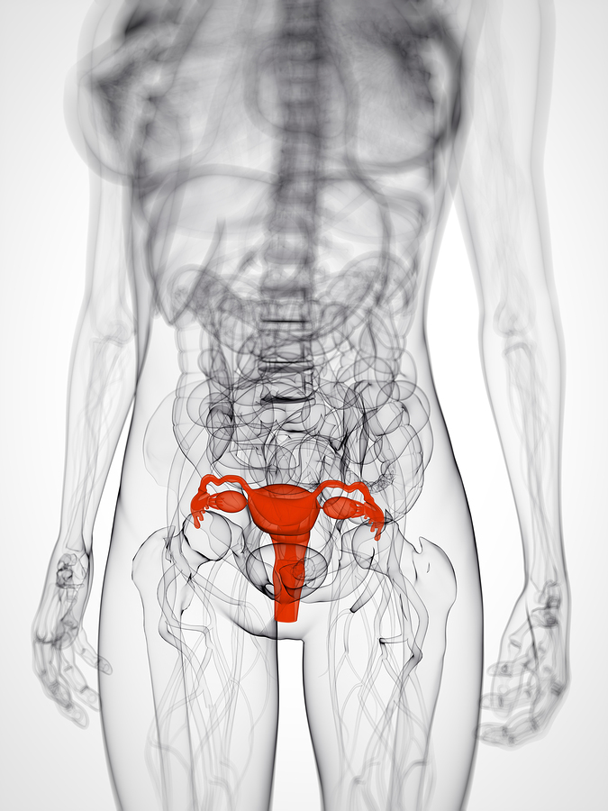
Staging Of Cancer Of The Uterus
Tumor growth behavior
The lining of the uterus (called “endometrium” by doctors) goes through similar cell changes in the development of cancer, as is the case in cervical cancer, where I described this process in detail.
The equivalent for “dysplasia” in cancer of the cervix would be called “atypical adenomatous hyperplasia” in uterine cancer. I would not want you to remember that, but understand the principal that there are warning signs that tell the gynecologist, when an endometrial biopsy is done, that this type of change will go on to develop into uterine cancer. Since the early 1990’s endometrial biopsies have become a more common procedure and this can significantly determine the risk for uterine cancer development down the road (see below).
Metastases of cancer of the uterus
Once cancer of the uterus is established, it has the tendency to grow along the uterine cavity down into the cervix and also towards the fallopian tubes. The cancer cells produce lytic enzymes, which enable the cancer also to eat its way slowly through the uterine wall. Frequently the cancer invades the ovaries, the uterine support ligaments (called “broad ligament”), the vagina and the pelvic lymph nodes. Next the cancer cells spread further through the lymphatic passages into the paraaortic lymph nodes and into the blood stream.
From here metastases can reach any organ. Inside the abdomen the cancer cells shed into the peritoneal cavity from either the fallopian tube, an invasion through the uterus or through an ovarian metastasis. The cells can also migrate through lymphatic migration channels (described in more detail in the ovarian cancer chapter) into the liver, the right lung or into retroperitoneal lymph glands.
Round ligament and pelvic lymph gland metastases
There is another channel for metastases to migrate through and this is along the round ligaments (a uterine ligamentous support to the front of the lower abdominal wall) and through lymphatic connections to the thigh lymph nodes (called” femoral nodes”). If there are lumps under the skin of the upper front thigh region in a woman with uterine cancer, this is a sign of a very poor survival rate.
It can be difficult to establish the stage of uterine cancer. Sometimes it appears that the disease only affects 10% of the pelvic lymph glands. However, autopsy studies often show later that much more invasive disease was present with metastases in 65% of the pelvic lymph glands (Ref. 1, p. 1196).
References
1. Cancer: Principles &Practice of Oncology.4th edition. Edited by Vincent T. DeVita, Jr. et al. Lippincott, Philadelphia,PA, 1993. Vol. 1. Chapter on gynecological tumors.
2. Cancer: Principles&Practice of Oncology. 5th edition, volume 1. Edited by Vincent T. DeVita, Jr. et al. Lippincott-Raven Publ., Philadelphia,PA, 1997. Chapter on gynecological tumors.
3. B. Sears: “The age-free zone”. Regan Books, Harper Collins, 2000.
4. E Weiderpass et al. Cancer Causes Control 2000 Feb;11(2):185-192.
5. S Shibutani et al. Cancer Res 2001 May 15;61(10):3925-3931.
6. DB Fournier et al. Gynecol Oncol 2001 Jun;81(3):366-372.
7. DS McMeekin et al. Gynecol Oncol 2001 May;81(2):273-278.
8. LA Katz et al. Am J Obstet Gynecol 2001 May;184(6):1071-1073.
9. B Bonanni et al. Breast J 2000 Oct;6(5):317-323.
10. MG Jain et al. Eur J Epidemiol 2000;16(10):899-905.
11. Conn’s Current Therapy 2004, 56th ed., Copyright © 2004 Elsevier
12. Ferri: Ferri’s Clinical Advisor: Instant Diagnosis and Treatment, 2004 ed., Copyright © 2004 Mosby, Inc


