Introduction
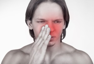
Sinusitis exists when one or more of the sinus cavities is infected. There are 3 pairs of common sinus cavities, the frontal sinuses above the eye sockets, the ethmoid sinuses between the nasal cavity and the eye sockets, and the maxillary sinuses underneath the eye sockets, but above the upper row of teeth.
There is a fourth location of a sinus cavity, located in the midline right underneath the pituitary gland, which is called the sphenoid sinus. These hidden cavities in the facial bone are lined with a mucous membrane and are connected to the inside of the nose through very tiny ducts.
Blocking of sinus ducts and development of sinusitis
These can get plugged with a cold, which can lead to sinusitis with a “sinus headache”. Subsequent bacterial superinfection can lead to an acute bacterial sinusitis. Often the pathogen is a bacterium such as Haemophilus influenzae or Staphylococcus aureus, but viruses can also cause an identical clinical picture. As the sinus ducts are plugged and a vacuum develops inside the sinus cavities, there is an accumulation of inflammatory serum, which is the ideal breeding ground for bacteria to multiply in.
Symptoms
With regard to sinus symptoms, there may be a dull pain around the eyes, there may be a pussy discharge from the nose and a fever. Depending on which sinuses are affected, there can be swelling over the area.
For instance, with maxillary sinusitis there might be swelling and tenderness in the area below the side of the nose underneath the eye socket. At the same time there might be a tooth ache in the upper teeth as the nerve roots can be directly irritated from inflammation in the bottom part of the sinus cavity where the nerves run by. With a frontal sinusitis there is often a frontal headache. With ethmoid sinusitis there is a splitting headache in he front and pain between and behind the eyes. A sphenoid sinusitis gives the patient a more dull headache either in the back or in the front.
Diagnostic tests
In an acute sinusitis the doctor may make the diagnosis clinically and treat with a course of antibiotics. In chronic sinusitis, which has the identical symptoms as acute sinusitis, diagnostic tests may be necessary to locate the sinusitis and look for other underlying causes. A CT scan can give a lot of detail, shows the extend of the sinusitis, possible underlying polypoid or cancerous lesions etc. that may have predisposed the patient to get sinusitis.
Treatment
Sinus treatment consists of doing steam inhalation frequently and for 10 minutes at a time. This will bring the swelling of the nasal lining down facilitating the opening up of the sinus ducts and promoting drainage.
Topical vasoconstrictive nasal sprays such as phenylephrine (brand names: Dionephrine, Mydfrin, Neo-Synephrine) or xylometazoline nasal spray (brand names: Otrivin, Decongest) will also assist in drainage of sinus cavity secretions. However, the patient should not take these nasal solutions more than 7 days in a row as they lose effectiveness. In acute sinusitis the physician orders penicillin V or erythromycin or 10 to 12 days. With chronic sinusitis the patient receives amoxicillin or tetracycline as a prolonged course over 4 to 6 weeks. With pussy nasal discharge the physician takes a bacterial culture to detect the pathogen. This guides him about the choice of antibiotic.
Referral to ENT specialist
If the doctors sees that a chronic sinusitis does not respond to the above-mentioned measures, he may decide to refer to an ENT specialist. The specialist may decide to do a surgical drainage procedure with endoscopic intranasal surgery to ventilate the sinuses again. In immune deficient patients, such as AIDS patients or patients with poor control of diabetes or recipients of transplanted organs on immune suppressants, chronic sinusitis may develop with more rare fungal infections.
Mucormycosis is one such fungal infection, which leads to black dead tissue from which the lab technician can isolate fungus. It would need treatment with intravenous amphotericin B, an antifungal agent, and improvement of the diabetic control, if this is the underlying metabolic condition.
Aspergillosis common in cancer patients
The physician suspects aspergillosis in a person with cancer who is on chemotherapy. But a patient may have immune compromise when there is polypoid tissue in the nose and the sinuses. There are several species such as Aspergillus flavus, A.fumigatus and A.niger. The specialist needs to do a biopsy and culture of this material and once confirmed as aspergillosis, wide surgical drainage and cleaning out of the papillomatous material has to be done in combination with intravenous amphotericin B (brand name: Fungizone), which eradicates this fungus. If the seriousness of this condition is not appreciated, this disease can be fatal as it will spread systemically (Ref. 4, p.689).
Candidiasis in immunosuppressed patients
Candidiasis is common in AIDS patients and patients with immunosuppressive therapy or diseases. It is very versatile and causes white thrush on the mucous membranes of the mouth or genitals (glans of penis, inside vagina), or moist skin areas between the fingers, or in moist skin folds particularly in obese people. With regard to the sinuses the accumulation of mycel in the sinus ducts can lead to blockage of the natural drainage of the sinuses, which leads to the candidiasis infection. Treatment: Similar to aspergillosis the specialist needs to biopsy and culture the mycel material. When the physician diagnoses Candida albicans, the ENT specialist may have to do drainage procedures to reopen the sinuses wide. The physician combines this with anti Candida albicans therapy such as fluconazole (brand name: Diflucan) orally. The physician treats serious systemic infection with Amphotericin B (brand name: Fungizone) intravenously (Ref. 1, p. 83).
References
1. The Merck Manual, 7th edition, by M. H. Beers et al., Whitehouse Station, N.J., 1999. Chapter 161.
2. TC Dixon et al. N Engl J Med 1999 Sep 9;341(11):815-826.
3. F Charatan BMJ 2000 Oct 21;321(7267):980.
4. The Merck Manual, 7th edition, by M. H. Beers et al., Whitehouse Station, N.J., 1999. Chapter 43.
5. JR Zunt and CM Marra Neurol Clinics Vol.17, No.4,1999: 675-689.
6. The Merck Manual, 7th edition, by M. H. Beers et al., Whitehouse Station, N.J., 1999. Chapter 162.
7. LE Chapman : Antivir Ther 1999; 4(4): 211-19.
8. HW Cho: Vaccine 1999 Jun 4; 17(20-21): 2569-2575.
9. DO Freedman et al. Med Clinics N. Amer. Vol.83, No 4 (July 1999): 865-883.
10. SP Fisher-Hoch et al. J Virol 2000 Aug; 74(15): 6777-6783.
11. Mandell: Principles and Practice of Infectious Diseases, 5th ed., copyright 2000, Churchill Livingstone, Inc.
12. Goldman: Cecil Textbook of Medicine, 21st ed., Copyright 2000, W. B. Saunders Company
13. PE Sax: Infect DisClinics of N America Vol.15, No 2 (June 2001): 433-455.
14. Suzanne Somers: “Breakthrough” Eight Steps to Wellness– Life-altering Secrets from Today’s Cutting-edge Doctors”, Crown Publishers, 2008


