With regard to treatment of squamous cell cancer it is absolutely important for the patient to act, as we know that squamous cell cancer tends to metastasize early and any delay of treatment leads to a higher mortality.
The doctor can only help those patients who come to see him/her. In the past, particularly in Germany in the early 1970’s I have seen some terribly advanced cases of this type of cancer. It has become more and more apparent to the medical profession and fortunately also now to the general population how important it is to diagnose skin cancer early.
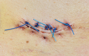
There are a few weeks, perhaps even months, where squamous cell cancer grows locally. But the larger the cancer lesion is, the more likely that some cancer cells are shed into the lymphatic drainage and from there into the blood circulation. I cannot overemphasize therefore how urgent it is to get the suspicious skin lesion removed, before this spread of cancer cells occurs. There are many skin lesions that are harmless. But there are others that may look harmless to a lay person, but that actually are disguised squamous cell cancers. I have been surprised many times how a harmless looking skin lesion that I removed (“just to be safe”) turned out to be an early squamous cell cancer. Often the skin biopsy, where the physician will remove the smaller lesion completely in the healthy tissue, is at the same time the appropriate treatment for this condition.
The pathologist usually looks at all the margins to make sure that the whole cancer is removed. Some of the more widespread squamous cell cancers need to be seen by a dermatologist and often require the Mohs’ micrographic surgery mentioned under the basal cell carcinoma to ensure complete excision. After the surgery has been successfully done close regular follow-up visit protocol should be followed for a period of time to be specified by the treating surgeon to ensure early diagnosis of recurrences. The unfortunate patients that I remember from Germany in the early 1970’s failed on both early diagnosis and the close post surgical follow-up visits.
Sometimes it might be suggested that the dermatologist observe a skin lesion and photographs may be taken with a scale on it and this can be reassessed a few weeks or months later to document either growth or no growth. Any biopsy of a skin lesion must be sent for pathology. This allows for an accurate tissue diagnosis.
Radiation treatment has a place in areas of the body where surgery would be disfiguring such as on the nose, lip, nasolabial fold, around the eye and eyelid. Sometimes such radiation is easier for older patients, but for younger ones one has to take into consideration that one of the long-term side effects can be the development of skin cancer, the very thing we wanted to get rid of at the start. If at all possible one should therefore use the Mohs’ technique or plastic surgery methods to remove the cancer.
References:
1. Kripke ML, Sass ER,eds.”Antigenicity of murine skin tumors induced by UV light”.JNCI 1974;53:1333-1336.
2. Kripke ML. “Immunology and photocarcinogenenis”. J. Am. Acad.Dermatol. 1986;14:149-155.
3. Potter M.”Percivall Pott’s contribution to cancer research”. NCI Monogr.1963;10:1.
4. COMS Group: Collaborative ocular melanoma study manual of procedures. National Technical Information Service. Springfield,VA, 1989.
5. Cancer: Principles &Practice of Oncology. 4th edition. Edited by Vincent T. DeVita, Jr. et al. Lippincott, Philadelphia,PA, 1993. Chapter on Cancer of the skin.
6. Cancer: Principles&Practice of Oncology. 5th edition, volume 2. Edited by Vincent T. DeVita, Jr. et al. Lippincott-Raven Publ., Philadelphia,PA, 1997. Chapter on Cancer of the skin.
7. Conn’s Current Therapy 2004, 56th ed., Copyright © 2004 Elsevier
8. Ferri: Ferri’s Clinical Advisor: Instant Diagnosis and Treatment, 2004 ed., Copyright © 2004 Mosby, Inc
9. Suzanne Somers: “Breakthrough” Eight Steps to Wellness– Life-altering Secrets from Today’s Cutting-edge Doctors”, Crown Publishers, 2008


