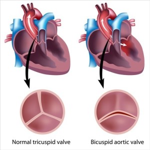
Aortic valve disease can develop as a result of a prior infection of the heart valves, called endocarditis. The aortic valve is located between the left heart chamber and the aorta.
The valve opens when the heart contracts (in systole) and closes between heart beats (in diastole) as shown here. The aortic valve may get narrowed (stenotic) due to endocarditis. Arteriosclerosis (hardening of the artery) from aging of the aorta can also affect the aortic valve and lead to a leaky valve (aortic insufficiency). About 2% of the population has a valve deformity where instead of the normal tricuspid (three leaflets) aortic valve there is a bicuspid (two leaflets) aortic valve. However, this is often only symptomatic in adult life when the bicuspid valve calcifies and leads to aortic insufficiency (see image).
Symptoms of patients with heart valve deformities
These patients may present with tiredness or some breathlessness with exercise or when walking stairs. This could go on for many years until suddenly one day the balance tips and they become acutely symptomatic. Now they present with symptoms such as air hunger, left sided chest pains and dizziness.
The skin color may be pale. The physician examining the patient will detect pathological heart sounds and murmurs on auscultation which assists with all the other signs and symptoms to lead to a diagnosis. A referral to a cardiologist will be the next step and further tests ordered by the specialist such as an echocardiogram (which are Doppler studies of the structures of the heart), X rays and possibly heart catheterization with angiocardiography will pinpoint the exact diagnosis.
Examination of patient by cardiologist
Following this the cardiologist can formulate a rational treatment plan involving either surgery, medication or a combination of both.
The two major conditions that may be found are either an aortic insufficiency where the valves of the aorta do not close properly and blood regurgitates into the heart between two heart beats (during diastole) or an aortic stenosis where the valves are scarred and will not open enough during the heart beat (called systole). In the last few years much has been gained in this field and even heart transplantation has become a feasible alternative for the end stage of CHF. In the past this meant almost always certain impending death. Here is a story about a happy outcome with surgery.
Other Heart Valves
There are two more heart valves in the right heart chamber (the tricuspid and the pulmonary valves) that can also get diseased. These abnormalities are much less common. Congenital heart valve abnormalities are not uncommon including variations of heart valves such as the mitral valve prolapse and other defects. These are beyond the scope of this website. However, the family physician can do an initial assessment and refer the affected patient to the appropriate specialist.
References
1. The Merck Manual, 7th edition, by M. H. Beers et al., Whitehouse Station, N.J., 1999. Chapters 197, 202, 205 and 207.
2. Braunwald: Heart Disease: A Textbook of Cardiovascular Medicine, 6th ed., 2001, W. B. Saunders Co.
3. D C Bauer: Audio-Digest Family Practice Vol. 49, Iss. 09, March 2, 2001.
4. Ferri: Ferri’s Clinical Advisor: Instant Diagnosis and Treatment, 2004 ed., Copyright © 2004 Mosby, Inc.
5. Rakel: Conn’s Current Therapy 2004, 56th ed., Copyright © 2004 Elsevier


