Introduction
To clarify, what exactly is an aortic aneurysm? An aortic aneurysm is a bulging out of the normal caliber of the aorta, the main artery coming from the heart and going down along the spine just behind the back of the abdominal wall. To emphasize, two main risks are smoking and high blood pressure, both of which accelerate arteriosclerosis and lead to a softening of the wall of the aorta.
To explain, as the aortic wall gets soft with a loss of supportive elastic fibers and a breakdown of the lining, a bubble of the diseased aortic section forms, which at one point ruptures suddenly. In general, most of the aortic aneurysms form in the aortic section underneath the level where the kidney arteries branch off and the bifurcation into the iliac arteries at the pelvic floor.
Symptoms
For one thing, the cardinal symptom for a developing aortic aneurysm is abdominal pain. Specifically, as the aorta is situated so far in the back, the piercing deep pain is often felt in the lower back region and might be mistaken for lumbosacral, musculoskeletal pain.
Truly, there may be a new pulsating feeling deep in the mid abdomen. Indeed, often the physician can gently verify through palpation that this actually is so. Certainly, the pain tends to be steady and the location of the pain is dictated by the location of the aneurysm. For example, it would be in the central region of the abdomen, but can be in the epigastric region (upper mid abdomen), in the mid abdomen or in the suprapubic area (lower mid abdomen). By all means, it is important both for the physician and the patient not to rest until the cause of the pain has been identified. In fact, this might be the patient’s only chance for survival as a ruptured aorta has a high mortality rate through internal bleeding.
Treatment
For one thing, the key investigation is to order an abdominal ultrasound right away. Notably, it can help in sizing the aneurysm. To be sure, occasionally the physician orders a CT scan or MRI scan in addition, but often this is not necessary. Certainly, what is necessary is that a cardiovascular surgeon assesses the patient.
In this case, the specialist may order abdominal aortography, an X-ray procedure where a catheter is introduced through a needle into the femoral artery and advanced into the aorta.
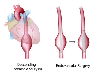
Frequently a contrast material can give a clear picture of the exact formation of the aneurysm. It is important to realize the after this the cardiovascular surgeon will then know exactly what section needs to be removed. Surgery on an emergency basis for an aortic dissection or ruptured aneurysm has a mortality risk of 50%. However, if the patient reports the early symptoms of an aneurysm earlier, the patient’s mortality risk from an elective surgical procedure to repair the aortic aneurysm is only 2 to 4 %. Here is a link to a site with images and information about surgery for aortic aneurysms. There are newer endoscopic procedures where the Seldinger technique is used to place a stent to overbridge the aneurysm.
References
1. DM Thompson: The 46th Annual St. Paul’s Hospital CME Conference for Primary Physicians, Nov. 14-17, 2000, Vancouver/B.C./Canada
2. C Ritenbaugh Curr Oncol Rep 2000 May 2(3): 225-233.
3. PA Totten et al. J Infect Dis 2001 Jan 183(2): 269-276.
4. M Ohkawa et al. Br J Urol 1993 Dec 72(6):918-921.
5. Textbook of Primary Care Medicine, 3rd ed., Copyright © 2001 Mosby, Inc., pages 976-983: “Chapter 107 – Acute Abdomen and Common Surgical Abdominal Problems”.
6. Marx: Rosen’s Emergency Medicine: Concepts and Clinical Practice, 5th ed., Copyright © 2002 Mosby, Inc. , p. 185:”Abdominal pain”.
7. Feldman: Sleisenger & Fordtran’s Gastrointestinal and Liver Disease, 7th ed., Copyright © 2002 Elsevier, p. 71: “Chapter 4 – Abdominal Pain, Including the Acute Abdomen”.
8. Ferri: Ferri’s Clinical Advisor: Instant Diagnosis and Treatment, 2004 ed., Copyright © 2004 Mosby, Inc.
9. Suzanne Somers: “Breakthrough” Eight Steps to Wellness– Life-altering Secrets from Today’s Cutting-edge Doctors”, Crown Publishers, 2008


