Introduction
First of all, cholecystitis is usually an inflammation of the gall bladder wall, often without any infection. It is important to realize that it comes from the closing of the cystic duct. This can lead to swelling of the gall bladder wall and fluid in the surrounding area (inflammatory exudate).
The word “cholecystitis” is doctor language, but is easy to understand: the first part”chole-” means “gall”, the second part “cystitis” means “bladder inflammation” or “bladder infection”. In this case it would mean “gall bladder inflammation”.
Gall bladder symptoms
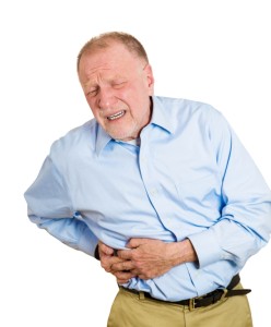
In the first place, symptoms are very similar to the symptoms described under cholelithiasis (gall stones). Furthermore, there is evidence the two travel side by side. In like manner, it appears that cholecystitis is caused from the presence of gall stones or sludge. This can lead to intermittent obstruction of the cystic duct, in the meantime. A typical attack would start in due time with a colicky pain (biliary colic) following a fatty meal. At the same time, the pain is severe and nausea and vomiting may also occur. This pain typically locates in the right upper abdomen. But it can travel to the right lower shoulder blade. Probably the pain then subsides in about 2 or 3 days and is often gone in 1 week. As a matter of fact, during the peak of the pain the patient likely is hospitalized and tests are performed.
Diagnosis
Cholescintigraphy (gallbladder scan) is done without delay with intravenous iminodiacetic acid compounds that are labeled with radioactive technetium 99m. This compound is metabolized by the liver and sent through the bile ducts into the gall bladder and the duodenum.
However, with acute cholecystitis the gall bladder is not visualized, whereas the liver and bile ducts are normal. To summarize, this would be a positive test to indicate the presence of acute cholecystitis. In addition, ultrasound (=sonography) studies show the thickening of the gall bladder wall. There is also fluid in the surrounding area of the gall bladder (inflammatory exudate).
The differential diagnosis (= considering alternative diagnoses) of cholecystitis should include such things as gangrene and perforation of the gall bladder. This is a life threatening condition, where peritonitis ensues. A surgeon does a laparotomy immediately. Acute acalculous cholecystitis (=cholecystitis without the presence of gall stones) is another serious illness. It is treated by intravenous feeding of the patient and cholecystectomy as soon as this can be done safely.
Treatment
As shown above, the physician is hospitalizing a patient with cholecystitis and and treating with intravenous rehydration. In any event, there is no oral feeding and a nasogastric tube removes all of the digestive secretions. Overall, if the clinical presentation and the blood tests suggest infection, the physician administers intravenous antibiotics. As said before, the patient usually settles within 3 days. The surgeon will decide whether to do an early or a delayed cholecystectomy. In essence, this depends on the clinical presentation.
Chronic cholecystitis
This condition is associated with a chronically diseased gall bladder. The gall bladder wall is thick and fibrotic and it may contain sludge or gall stones. The patients may have had the typical gall stone symptoms. But they may now experience them for the first time. The common denominator is that all these patients have evidence of chronic gall bladder disease. They need gall bladder surgery to stop the gall bladder attacks. Many patients suffer quietly and try to remedy their symptoms with a”gall bladder diet”. However, these patients need a laparoscopic gall bladder surgery (cholecystectomy). Then they become symptom free. It still makes sense to go on a low fat diet. It also makes sense to aim for a normal weight. And it is advisable to regulate the bowel habits by high fiber food intake (salads, vegetables, full grain products).
References
1. M Frevel Aliment Pharmacol Ther 2000 Sep (9): 1151-1157.
2. M Candelli et al. Panminerva Med 2000 Mar 42(1): 55-59.
3. LA Thomas et al. Gastroenterology 2000 Sep 119(3): 806-815.
4. R Tritapepe et al. Panminerva Med 1999 Sep 41(3): 243-246.
5. The Merck Manual, 7th edition, by M. H. Beers et al., Whitehouse Station, N.J., 1999. Chapters 20,23, 26.
6. EJ Simchuk et al. Am J Surg 2000 May 179(5):352-355.
7. G Uomo et al. Ann Ital Chir 2000 Jan/Feb 71(1): 17-21.
8. PG Lankisch et al. Int J Pancreatol 1999 Dec 26(3): 131-136.
9. HB Cook et al. J Gastroenterol Hepatol 2000 Sep 15(9): 1032-1036.
10. W Dickey et al. Am J Gastroenterol 2000 March 95(3): 712-714.
11. M Hummel et al. Diabetologia 2000 Aug 43(8): 1005-1011.
12. DG Bowen et al. Dig Dis Sci 2000 Sep 45(9):1810-1813.
13. The Merck Manual, 7th edition, by M. H. Beers et al., Whitehouse Station, N.J., 1999.Chapter 31, page 311.
14. O Punyabati et al. Indian J Gastroenterol 2000 Jul/Sep 19(3):122-125.
15. S Blomhoff et al. Dig Dis Sci 2000 Jun 45(6): 1160-1165.
16. M Camilleri et al. J Am Geriatr Soc 2000 Sep 48(9):1142-1150.
More references
17. MJ Smith et al. J R Coll Physicians Lond 2000 Sep/Oct 34(5): 448-451.
18. YA Saito et al. Am J Gastroenterol 2000 Oct 95(10): 2816-2824.
19. M Camilleri Am J Med 1999 Nov 107(5A): 27S-32S.
20. CM Prather et al. Gastroenterology 2000 Mar 118(3): 463-468.
21. MJ Farthing : Baillieres Best Pract Res Clin Gastroenterol 1999 Oct 13(3): 461-471.
22. D Heresbach et al. Eur Cytokine Netw 1999 Mar 10(1): 7-15.
23. BE Sands et al. Gastroenterology 1999 Jul 117(1):58-64.
24. B Greenwood-Van Meerveld et al.Lab invest 2000 Aug 80(8):1269-1280.
25. GR Hill et al. Blood 2000 May 1;95(9): 2754-2759.
26. RB Stein et al. Drug Saf 2000 Nov 23(5):429-448.
27. JM Wagner et al. JAMA 1996 Nov 20;276 (19): 1589-1594.
28. James Chin, M.D. Control of Communicable Diseases Manual. 17th ed., American Public Health Association, 2000.
29. The Merck Manual, 7th edition, by M. H. Beers et al., Whitehouse Station, N.J., 1999. Chapter 157, page1181.
30. Textbook of Primary Care Medicine, 3rd ed., Copyright © 2001 Mosby, Inc., pages 976-983: “Chapter 107 – Acute Abdomen and Common Surgical Abdominal Problems”.
31. Marx: Rosen’s Emergency Medicine: Concepts and Clinical Practice, 5th ed., Copyright © 2002 Mosby, Inc. , p. 185:”Abdominal pain”.
32. Feldman: Sleisenger & Fordtran’s Gastrointestinal and Liver Disease, 7th ed., Copyright © 2002 Elsevier, p. 71: “Chapter 4 – Abdominal Pain, Including the Acute Abdomen”.
33. Ferri: Ferri’s Clinical Advisor: Instant Diagnosis and Treatment, 2004 ed., Copyright © 2004 Mosby, Inc.
34. Suzanne Somers: “Breakthrough” Eight Steps to Wellness– Life-altering Secrets from Today’s Cutting-edge Doctors”, Crown Publishers, 2008


