
Introduction
In the first place, like with the treatment of cancer of the cervix the physician needs to plan treatment of cancer of the uterus carefully. In particular, he needs to take into consideration the exact location of the cancer at the time of diagnosis.
The gynecologist/oncologist will include information about the histology, the grade of the tumor and the cancer stage before the ideal treatment modality and/or combination can be recommended for the patient. Certainly, a large body of literature exists from many clinical trials. Because of this treatments and survivals are now about 15 to 20% better for most stages of the cancer in comparison to the early 1980’s.
Five-year survivals for Uterine Cancer
In fact, one such study involved a group of patients with uterine cancer where researchers separated them into grades and these women received surgical treatment in combination with or without radiation.
The following 5-year survivals resulted from this (data averaged from Ref. 1, p. 1198 and 1200, arranged as a table here).
Vaginal recurrence rate (different grades of uterine cancer)
| Grade: | With radiation: | Without radiation: |
| 1 | 4% | 4% |
| 2 | 3% | 9% |
| 3 | 9% | 24% |
It is clear from this that there was a 15% survival advantage for grade 3 tumors with radiation therapy of the pelvic lymph nodes. Notably, radiation neutralized the high number of invisible lymph gland metastases in grade 3 tumors and improved survival.
Similarly, there were studies regarding recurrence rates after hysterectomy for grade 1 patients. They only received surgery. For example, in one such study researchers followed patients who developed isolated high vaginal recurrences. In this case they treated them with a combination of external beam radiation and brachytherapy (see below) and then followed them for 5-year survival studies. Notably, the tumor size at the time of the recurrence determined the longterm survival. In this case the table shows that there was a much better survival with the smaller lesions than with larger lesions. Certainly, both groups received radiotherapy. In this case oncologists incorporated these type of observations into the present day treatment against uterine cancer.
5-year survival for isolated high vaginal recurrence (uterine cancer)
| size of lesion (diameter): | % survival: |
| 2 cm or less | 74% |
| more than 2 cm | 30% |
By all means, treatment against uterine cancer has become complex as so many variables have entered into the equation. Most specialists treat early cancer like stage IA and IB with surgery only, although some authorities err on the cautious side and cover with some form of radiotherapy.
As the table below shows, stage IC and stage II do best with a combination of surgery and radiotherapy. Specialists treat stage III and IV usually with radiotherapy alone because surgery or combination surgery and radiotherapy did not yield better survivals. Specifically, there is a subgroup of patients with stage IV disease who will benefit from a radical surgical procedure called ” pelvic exenteration”. They have to be in fairly stable condition for this surgical procedure, in which all of the visible cancerous tissue is removed in an extensive debulking procedure. Subsequent to that radiotherapy and chemotherapy to prevent recurrence is given. 5- year cure rates of 40% to 50% have been achieved this way!
5-year survival for uterine cancer
| Stage (FIGO): | % survival: | Comments: |
| IA, IB | 90% | low risk: surgery |
| IC | 80% | higher risk: surgery+radio-Rx |
| II | 75% | Surgery + radiotherapy |
| III | 46% | extended field radio-Rx for paraaortic lymph nodes |
| IV | 5%(or 40%*) |
| * With radical surgery (called “pelvic exenteration”), followed by radiotherapy and chemotherapy, 5-year survivals of 40% to 50% have been reported, but not every patient is a suitable candidate for this very extensive surgery. |
Surgery
With this in mind, surgery consists usually of an abdominal hysterectomy and removal of both fallopian tubes and ovaries (called”bilateral salpingo-oophorectomy”). In addition, the oncological gynecologist also removes samples of pelvic and paraaortic lymph glands. All of the material is sent to the pathologist who will report back, which of the tissues contained cancer cells. This data is needed in order to decide whether radiotherapy is needed.
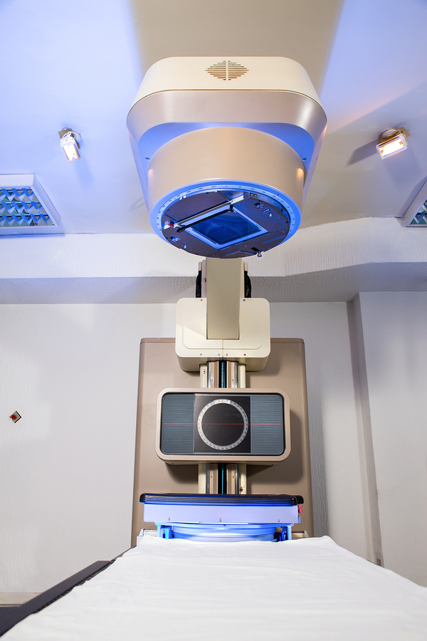
Treatment Of Cancer Of The Uterus (External beam therapy)
Radiotherapy
For one thing, radiotherapy is delivered in two ways. External beam therapy delivers radiation through the skin in overlapping beams that are mostly concentrating in the uterus, pelvic and paraaortic lymph gland areas.
Notably, using spatial computer models radiation doses can be accurately delivered in differently shaped pelvic cavities. and can be modified according to body size and shape. Equally important, this is supplemented by a direct dose of radiation from local “brachytherapy“. With this form of radiotherapy a device, which has an “arm” (the name “brachy” means arm) that fits into the uterus and another piece that fits into the upper vagina, is placed for several hours until a certain dose has been delivered to the tissues.
That is to say, between these two delivery methods the radiation specialist can deliver curative doses of radiation with a minimum of side effects. The older technique with radioactive radium has largely been replaced in the 1980’s with radioactive caesium (137-Cs), which has less side-effects than radium and is easier to apply. About 2% of patients experience major side effects such as bladder, rectum or gastrointestinal scarring. However , on the other hand this is considered a very acceptable risk given the alternatives of not treating the cancer.
Chemotherapy
It is important to realize that uterine cancer is not very sensitive to chemotherapeutic intervention. However, in spite of this multidrug combination therapy when combined with radiotherapy and surgery has made some difference in survival data as mentioned above. For one thing, there are still tremendous immunosuppressant side-effects that need to be overcome. For example, some future approaches might include monoclonal antibodies directed against tumor markers (Ref. 2).
References
1. Cancer: Principles &Practice of Oncology.4th edition. Edited by Vincent T. DeVita, Jr. et al. Lippincott, Philadelphia,PA, 1993. Vol. 1. Chapter on gynecological tumors.
2. Cancer: Principles&Practice of Oncology. 5th edition, volume 1. Edited by Vincent T. DeVita, Jr. et al. Lippincott-Raven Publ., Philadelphia,PA, 1997. Chapter on gynecological tumors.
3. B. Sears: “The age-free zone”. Regan Books, Harper Collins, 2000.
4. E Weiderpass et al. Cancer Causes Control 2000 Feb;11(2):185-192.
5. S Shibutani et al. Cancer Res 2001 May 15;61(10):3925-3931.
6. DB Fournier et al. Gynecol Oncol 2001 Jun;81(3):366-372.
7. DS McMeekin et al. Gynecol Oncol 2001 May;81(2):273-278.
8. LA Katz et al. Am J Obstet Gynecol 2001 May;184(6):1071-1073.
9. B Bonanni et al. Breast J 2000 Oct;6(5):317-323.
10. MG Jain et al. Eur J Epidemiol 2000;16(10):899-905.
11. Conn’s Current Therapy 2004, 56th ed., Copyright © 2004 Elsevier
12. Ferri: Ferri’s Clinical Advisor: Instant Diagnosis and Treatment, 2004 ed., Copyright © 2004 Mosby, Inc
13. Suzanne Somers: “Breakthrough” Eight Steps to Wellness– Life-altering Secrets from Today’s Cutting-edge Doctors”, Crown Publishers, 2008


